ABMI
Main menu:
- Home
- About ABMI
- Clinical Focus
- Monitoring Software
- TCD Systems
- Clinical Studies
- News/Investors
- Contact Us
Embedded Technologies
Monitoring Software > Off-Line Emboli Classification Software: Embol-STATION
The Embol - STATION® embed two innovative technologies:
1. EED® Technology (Enhanced Emboli Display)
Enhanced Emboli Display (EED®, patented) Technology provides caregivers with a new display for transcranial blood flow based on wavelet analysis. This new display greatly enhances embolic signals with respect to blood flow speckles, thus providing a much more visible, contrasted display of microembolic events. The numeric features drawn from the wavelets approach are successfully used for improving emboli detection - especially for low-power MES and for performing solid/gas differentiation.
EED® permits improved visualization of embolic events, especially in the case of small solids. On the illustration above, we display the raw Doppler signal (upper window), its FFT (middle window), and the EED® display (lower window). While the FFT is the reference display for blood flow, it turns out that EED visualization, based on wavelets analysis, shows embolic events with a much higher contrast, and reduces the saturation effect which usually results in impaired interpretation of the FFT display due to vertical bars.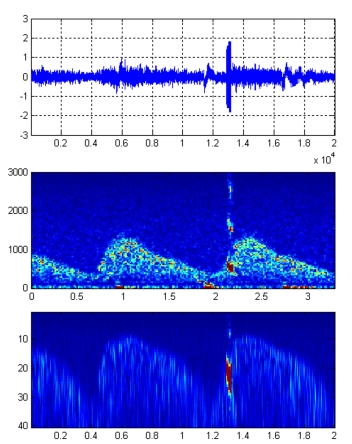
2. SSR® Technology (Saturated Signal Restoration)
Saturated signals often plague TCD recordings of embolic signals (especially for gaseous MES), rendering both Doppler and FFT displays filled with difficult-to-read vertical bars (harmonics). Saturated Signal Restoration Technology (SSR®, patented) permits signal restoration of the original, unclipped signal with good fidelity, thereby resulting in a more realistic display of the raw Doppler signal of the MES, as well as in a more readable FFT blood flow.
In addition, the subsequent differentiation of solid and gaseous MES improves when restored rather than saturated signals are used.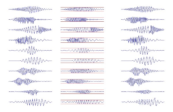
The above illustration shows 10 embolic signals on which we have tested SSR® technology. The first column displays10 original signals extracted from clinical patient monitorings. The 10 signals were then clipped or "cut" to 50% of their maximal amplitudes; the clipped signals are those displayed in column 2. Applying the SST signal restoration algorithm to the clipped signals allows for very good reconstruction of the 10 signals. The reconstructed signals are displayed in column 3, and it can be seen that they are indeed similar to the original, unclipped signals of column 1.